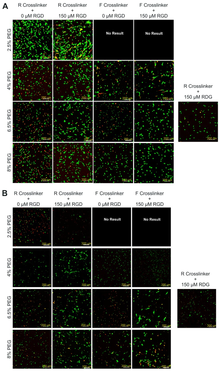
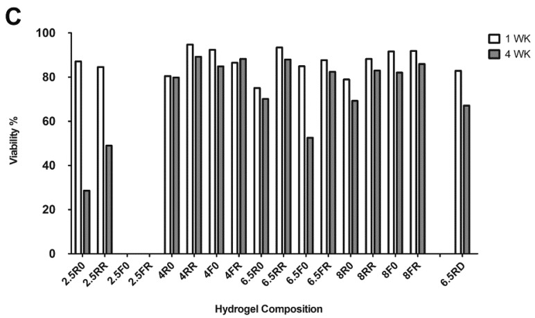
Figure 1.
Human periosteum-derived cell (hPDC) viability within PEG-VS hydrogels made with different concentrations of macromer, different protease sensitive cross-linkers (R or F), and with or without the cell binding motif RGD or scrambled RDG after 1 week (A) and 4 weeks (B) of in vitro culture in GM. Representative images of Live/Dead staining with calcein AM (green, live cells) and ethidium homodimer-1 (red, dead cells). Confocal images are depicted as the maximum intensity of a 500 μm z-stack that was acquired using a 10× objective (scale bars = 200 μm). (C) IMARIS software based quantification of viability percentages of cells encapsulated in polyethylene glycol (PEG) hydrogels (n = 1 independent sample; values depicted are the averages of two different measured areas within one sample).


