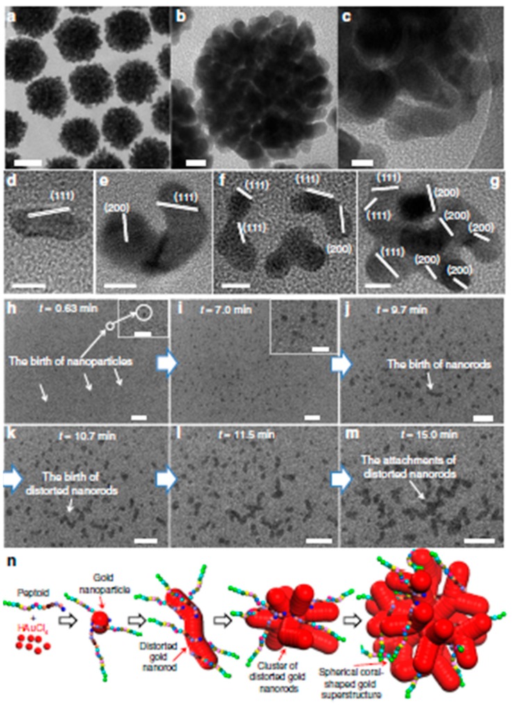
Figure 6.
(a,b) TEM images of the coral-shaped nanoparticles (scale bar; (a) 50 nm; (b) 10 nm). (c) HR-TEM (scale bar, 5.0 nm). (d–g) HR-TEM images of distorted gold nanorods and clusters in early stages of coral-shaped particle formation (scale bar, 5.0 nm). (h–m) Sequence of in situ liquid cell TEM time (scale bar, 20 nm) showing the early stages of coral-shaped particle formation (insets of h, i are the magnification of TEM images, scale bar at 10 nm). (n) Schematic representation for the multibranched formation of gold nanoparticles induced by peptoids. Reproduced from [51] with permission from the Nature Publisher.

