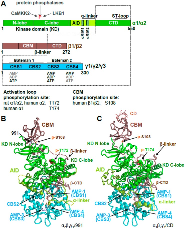
Figure 1.
Overall structure of human adenosine monophosphate (AMP)-activated protein kinase (AMPK). (A). Domain structure and AMPK isoforms. Activation loop and carbohydrate-binding module (CBM) phosphorylation sites of different isoforms are indicated below the domain map (B,C). Crystal structures of phosphorylated, AMP-bound AMPK α2β1γ1/991 ((B); PDB: 4CFE) and α1β2γ1/cyclodextrin (CD) ((C); PDB: 4RER).

