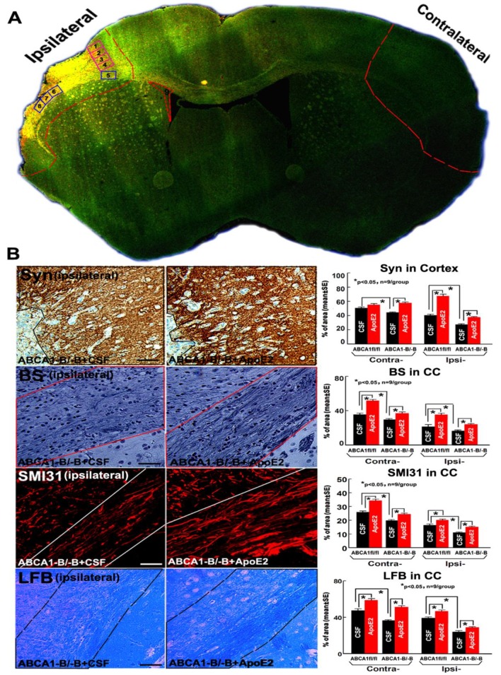
Figure 2.
Administration of ApoE2 increased brain grey matter (GM) and white matter (WM) densities in ABCA1−B/−B stroke mice 14 days after dMCAo: (A) Confocal-micrograph photo schematically shows the areas where the images were taken for Synaptophysin (Syn, square 1–4) and dendrite morphologies (square 2, 3) or Bielschowsky silver (BS)/Luxol Fast Blue (LFB)/phosphorylated high-molecular weight neurofilament (SMI31) (squares 5–8), nestin/Sox2 (square 9) and ischemic ipsilateral tissue and contralateral brain tissue (outlined areas); (B) Syn, BS, SMI31, and LFB staining and quantitative data. Scar bar = 40 µm.

