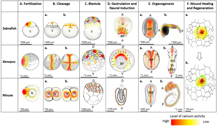
Figure 1.
Comparative calcium activity among zebrafish, frogs, and mice during egg activation and fertilization (A), first cleavage (Ba), second cleavage (Bb), blastula (C), gastrulation and neural induction (D) and organogenesis (E), including neural tube closure (Ea) and muscle development (Eb) in zebrafish, Xenopus, and mouse; also, depiction of calcium dynamics immediately after tissue damage (Fa) and calcium mediated actomyosin filament (green lining) during wound healing and regeneration (Fb). Black arrows show direction of propagation of calcium waves. Y, yolk; A, anterior; P, posterior; D, dorsal; V, ventral; BP, blastopore; NP, neural plate; UPM, unsegmented paraxial mesoderm; MS, matured somites; SS, segmenting somites; IS, injury site; N, nucleus. Images were adapted and re-drawn from [136,137,138].

