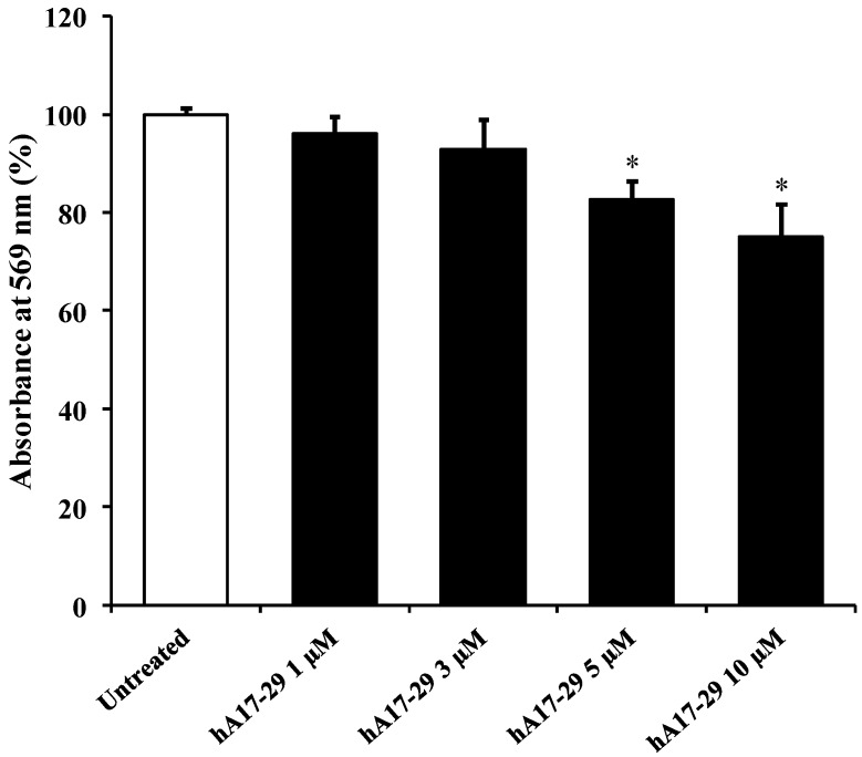
Figure 2.
Change in the cell viability caused by challenging for 48 h endothelial cells (RBE4) cells with different concentrations (1, 3, 5, and 10 µM) of freshly prepared hA17–29 peptide fragment. Cell viability was determined using the [3-(4,5-dimethylthiazol-2-yl)-2,5-diphenyltetrazolium bromide] MTT assay. MTT solution (1 mg/mL), obtained by dissolving the MTT powder in medium, was added to the cell cultures and incubated for 2 h at 37 °C; the formed crystals were melted with dimethylsulfoxide (DMSO) and used (200 µL of solution) to read the absorbance at 569 nm using a microplate reader (LabSystems-Multiskan Ascent 354 Microplate Reader, San Diego, CA, USA). Data are the mean of five independent experiments (an average of four readings was considered for each sample) and are expressed as the percent variation with respect to the absorbance at 569 nm recorded in untreated (control) cells. Standard deviations are represented by vertical bars. * Significantly different from untreated cells, p < 0.001.

