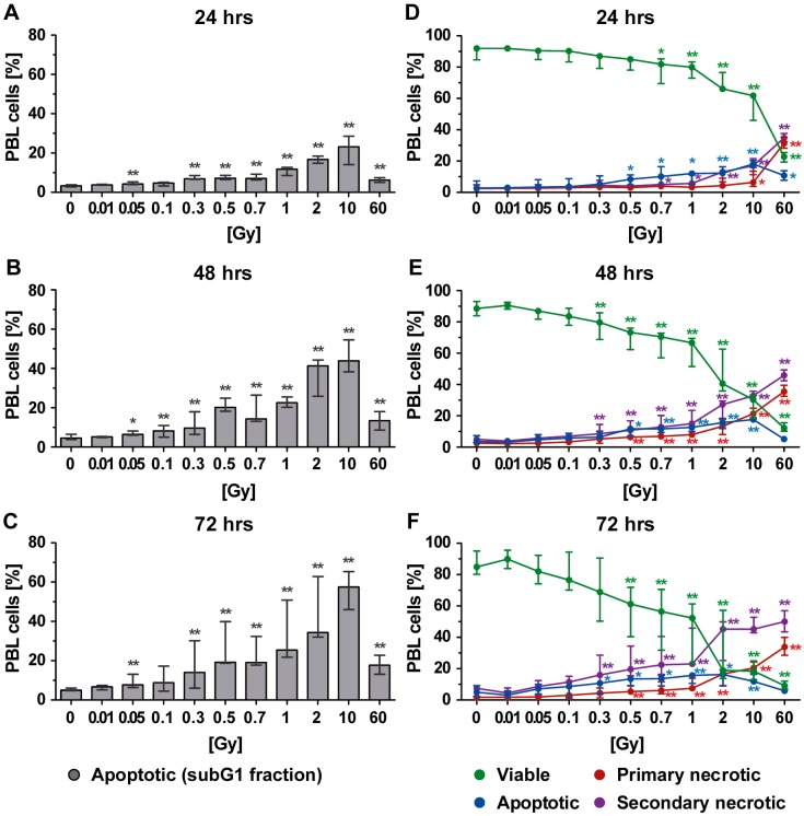
Figure 2.
Cell death in peripheral blood lymphoid cells (PBL) at different time points after irradiation as detected by analyses of (A–C) subG1 DNA content and (D–F) phosphatidylserine exposure and membrane permeability of PI with AxPI staining. Each data point represents the median (±interquartile range (IQR)) from six experiments from three different donors. Data points have been connected by lines to improve visual clarity. Statistical analyses were performed against the corresponding nonirradiated control (0 Gy) using the Mann–Whitney U test (* p < 0.05; ** p < 0.01).

