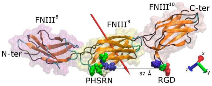
Figure 1.
Graphical representation of a FNIII8–10 fragment of fibronectin. The secondary structure (colour-coded; orange, teal, and black represent β-sheets, loops, and coil, respectively) along with the protein “ghost” surface of the modules that comprise the fragment are visible. The cell binding (RGD) and synergy (PHSRN) sites, as well as the distance between them, are shown, and the red arrow indicates the dipole moment of the fragment. The amino acids shown are colour-coded with blue, red, white, and green representing positively charged, negatively charged, hydrophobic, and polar uncharged, respectively. For sake of clarity, the water molecules are not shown.

