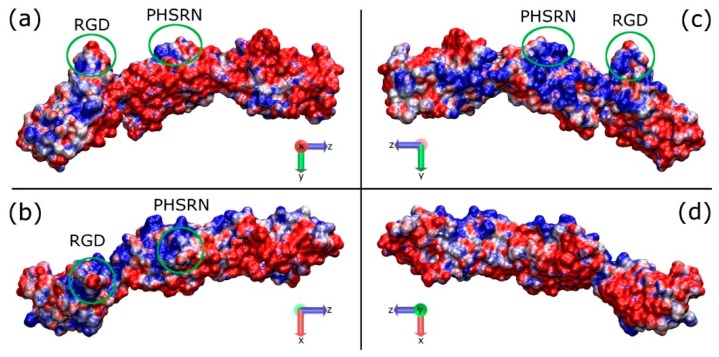
Figure 2.
Graphical representations of the FNIII8–10 domain showing the electrostatic properties (APBS) along the long axis of the protein; (a,c) the two sides of the protein on the y-z plane, (b,d) the two sides of the protein on the x-z plane. The sides from a to d are referred to as FNa, FNb, FNc, and FNd, respectively. The areas in the green circles indicate the RGD and PHSRN sites, which are facing up in (a) and (c), pointing out from the page in (b), and into the page in (d). Red and blue indicate the negatively and positively charged domains, respectively.

