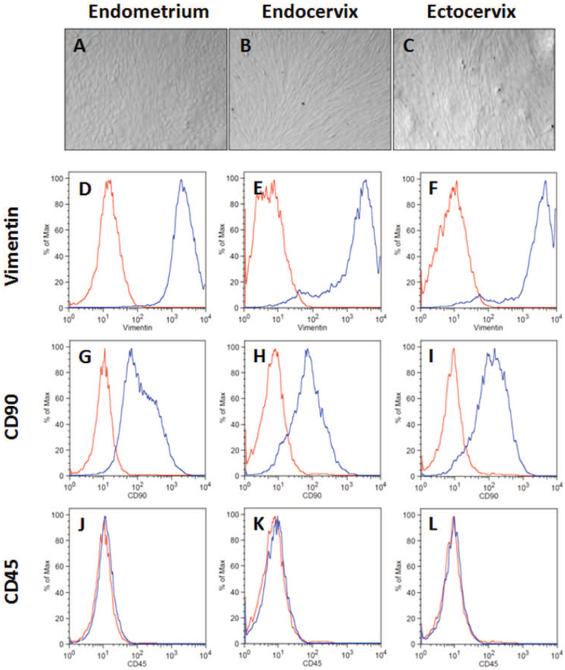
Figure 1: FRT stromal fibroblast morphology and marker expression.
Matched endometrial, endocervical, and ectocervical stromal fibroblasts were isolated from tissue fragments and cultured in vitro prior to analysis. (A-C) Representative images of unstained stromal fibroblasts imaged under a light microscope. Flow cytometry analysis of intracellular vimentin (D-F), surface CD90 (G-I) and surface CD45 (J-L) expression in matched endometrium, endocervix, and ectocervix from a representative individual donor. (Red – Isotype control; blue – antibody). All panels are derived from the same patient and are representative of 8 individual donors.

