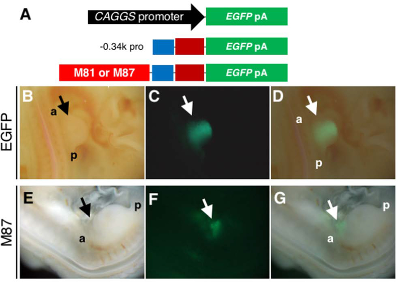
Figure 5. Activities of proximal promoter with and without two cis-elements in the developing chick limb bud.
(A) Schematics of constructs used in the Tol2 vectors. Top: the pCAGGS-EGFP. Middle: −0.34k promoter-EGFP. Bottom: −0.34k promoter-EGFP with either M81 sequence or M87 sequence.
(B-D) Images of the hindlimb bud of an embryo in which the Tol2-pCAGGS-EGFP and pCAGGS-transposase constructs were electroporated into the LPM of hindlimb-forming region. Bright field (B), EGFP (C) and a merged image (D) of the hindlimb bud. EGFP signal was observed throughout the limb mesenchyme at 48 hours after electroporation (n=11/15).
(E-G) Images of an embryo in which the Tol2–0.34k promoter-EGFP with the M87 sequence and pCAGGS-transposase constructs were electroporated into the LPM. Bright field (E), EGFP (F) and a merged image (G) of the hindlimb bud. Weak EGFP signal in the anterior edge of the hindlimb bud and the flank region at 48hr after electroporation.

