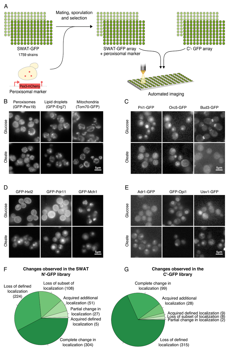
Figure 1. Unraveling the protein dynamics of yeast grown in oleate.
(A) A schematic diagram of the high content screen. Using an automated mating procedure we genomically integrated a peroxisomal marker, Pex3-mCherry to the SWAT N’ GFP library. Using a high-throughput fluorescence microscope, we screened this newly created library as well as the C’ GFP yeast library under growth in oleate-containing media. All images were taken at a single focal plane. We manually characterized the localization of each protein and compared it to its localization during growth on glucose-containing media. (B) Under growth in oleate more peroxisomes were observed, lipid droplets were larger and mitochondria were altered compared to growth in glucose. (C) Many changes that imply a general stress were observed in cells grown in oleate. Examples include loss of defined localization of Pri1, a DNA primase that is required for DNA synthesis and double-strand break repair, Orc5, a subunit of the origin recognition complex (ORC) which directs DNA replication and Bud3 that is involved in budding. (D) Many proteins were detected in the vacuole in oleate suggesting down regulation of various processes. Examples are Hxt2, a high affinity glucose transporter, and Pdr11, an ATP-binding cassette (ABC) transporter that moved from the plasma membrane to the vacuole. Mch1, a protein with similarity to mammalian monocarboxylate permeases that are involved in transport of monocarboxylic acids across the plasma membrane, moved from the vacuolar membrane to the vacuole. (E) Adr1, a transcription factor that regulates fatty acids utilization-related processes, Opi1, a negative transcriptional regulator of phospholipid biosynthetic genes, and Usv1 that affects transcriptional regulation of genes involved in growth on non-fermentable carbon sources, were localized to the nucleus under growth in oleate but not in glucose. (F) A pie chart summarizing the changes observed in the N’ GFP SWAT library. (G) A pie chart summarizing the changes observed in the C’ GFP library.

