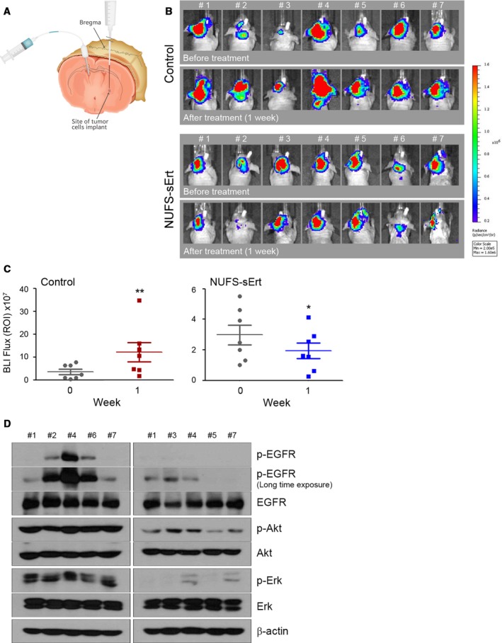
Figure 4.

Antitumor activity of NUFS‐sErt in an intracranial xenograft model. (A) Generation of the EGFR‐mutant lung adenocarcinoma intracranial model via intrathecal injections with HCC827‐Luc cells. Cells were injected into the left striatum, and the route of intrathecal injection was fixed into the right ventricle of the brain as described in the Materials and Methods section. (B, C) BLI images and quantification analysis of intracranial HCC827‐Luc tumor growth before and during treatment with NUFS‐sErt (20 μg/4 μL, 2 times/week, n = 7 animals) for 1 week. Red pseudo‐coloring indicates increased tumor growth, and green‐blue pseudo‐coloring indicates decreased tumor growth by bioluminescence quantification in B. (C) Quantification of the bioluminescence photon flux in the mice with intracranial HCC827‐Luc tumors treated over the indicated time points. Error bars are represented as mean ± SD. *P < 0.01 and **P < 0.001 by Student's t‐test. For all treatment studies, baseline imaging and subsequent therapy was initiated 14 days after intracranial tumor cell implantation. (D) Immunoblot analysis measuring each indicated pharmacodynamics biomarker in representative control‐treated or NUFS‐sErt‐treated tumors harvested from tumor‐bearing mice at 1 week following the initiation of therapy.
