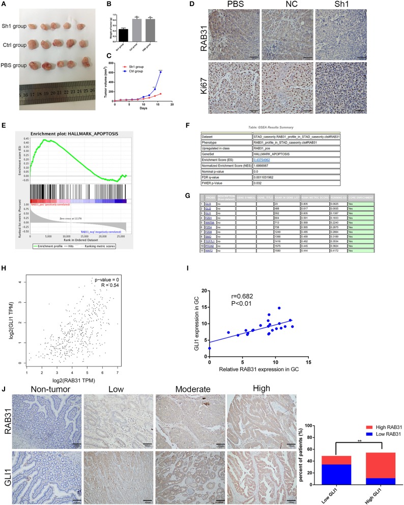
Figure 3.
Depletion of RAB31 suppressed tumor growth in vivo and biological data predicted RAB31 function via interaction with GLI1. (A) Xenograft tumors were excised from the mice. (B,C) Measurements of xenograft tumor volumes and weights. (D) IHC staining of RAB31 and Ki-67 in tumor specimens from mouse tumor tissues (Scale bar, 100 μm). (E) GSEA analysis of the TCGA dataset showed the association between RAB31 expression and apoptosis signaling pathways. The enrichment score (ES, green line) equals the degree to which the gene set is overrepresented. (G) Genes involved in apoptosis are listed according to rank metric score. (H) The relationship between RAB31 and GLI1 was determined using the GEPIA tool. (I) Fresh tissues from 22 pairs of patients were analyzed for a correlation between RAB31 and GLI1 mRNA levels by qPCR. (J) Association of RAB31 expression with GLI1 expression in 90 primary human GC specimens; the representative images are shown on the left (scale bar, 100 μm). The columns show the significant results on the right (**P < 0.01, ***P < 0.001).

