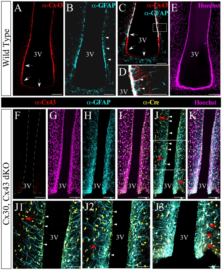
Figure 2.
Absence of hypothalamic Cx43 expression in double knockout (dKO) mice. Immunohistochemical analysis of frontal hypothalamic slices obtained from wild-type (WT; A–E) and dKO (F–J3) mice. The expression of Cx43 is shown in red, glial fibrillary acidic protein (GFAP) in cyan, and Cre recombinase in yellow; nuclei staining is shown in the magenta channel. Arrowheads in (B,D) indicate the boundary of GFAP expression and the co-localization of GFAP and Cx43 in the lateral wall of the 3V. Arrows in (A,C) show the expression of Cx43 in α-tanycytes and its absence or weaker expression in β-tanycytes. (F–K) Images of the ventricular area using the markers as described. (J1–J3) Images are higher magnifications of (J; white frames) from the dorsal to the ventral portion. Panels (J,J1,J2) indicate the expression of Cre recombinase in some tanycytes (arrowheads). Red arrows in the ventral region (J,J1–J3) point to the expression of Cre recombinase by astrocytes. 3V, third ventricle. Scale bar (A–K): 100 μm. Scale bar (J1–J3): 50 μm.

