
Figure 4.
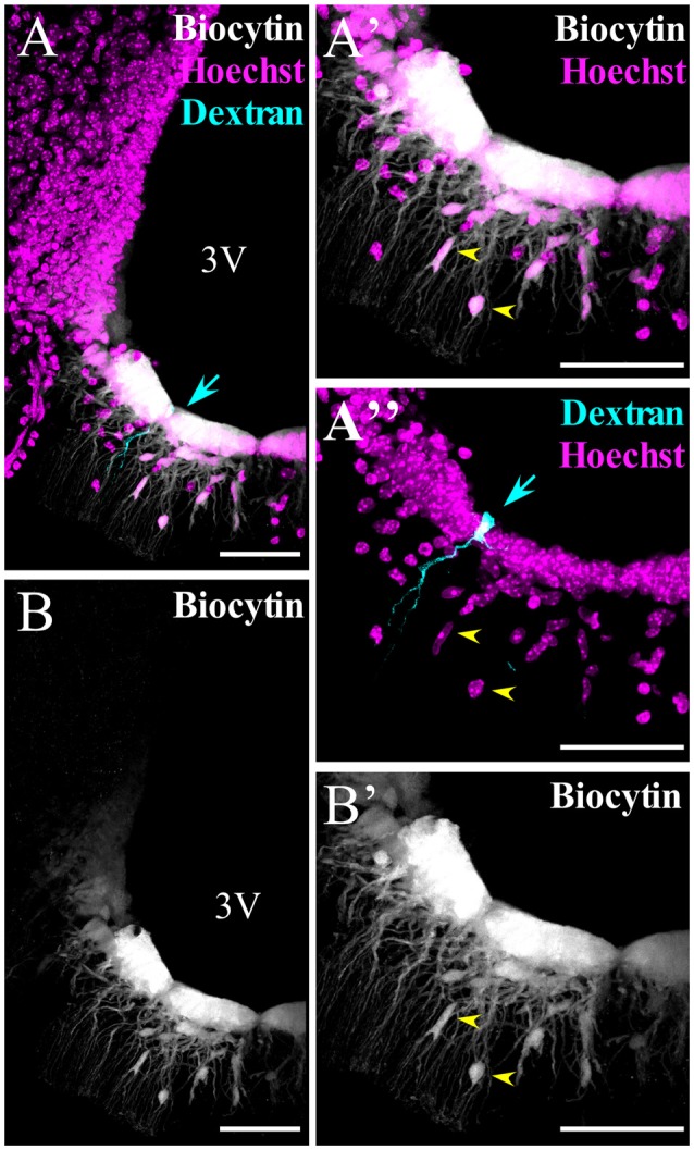
β-tanycytes are also highly coupled with each other and with parenchymal cells. (A–B′) Spread of biocytin through β-tanycytes and parenchymal cells. (A′,A″,B′) Higher magnification of the median eminence (ME) portion, where β-tanycytes localize. (A,A″) To identify the cell that was initially patched, it was filled with dextran Texas red (3 kDa, cyan arrow). Yellow arrows in (A′,A″,B′) show the parenchymal cells coupled. 3V, third ventricle. Scale bar: 50 μm.