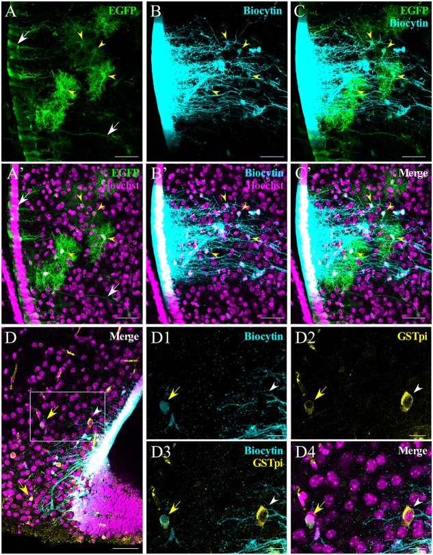
Figure 5.
Analysis of α-tanycyte-mediated panglial coupling. α-tanycytes in slices from human GFAP-enhanced green fluorescent protein (hGFAP-EGFP) mice were filled with biocytin to evaluate panglial coupling between tanycytes and astrocytes. (A) Immunohistochemistry against EGFP was performed in order to enhance the signal. White arrows indicate the somata and the processes of EGFP-labeled tanycytes. (B) Biocytin spreads to the neighboring tanycytes and parenchymal cells that co-localize with EGFP-positive astrocytes (C; yellow arrowheads). (A’–C’) show the respective images merged with the nuclear staining (Hoechst). (D–D4) Occasionally (in this case, in a Cx30 KO mouse), panglial coupling of the tanycyte with oligodendrocytes was observed. (D) Immunoreaction against GSTpi shows oligodendrocytes close to the ventral part of the hypothalamus. The merged pictures (D,D4) show GSTpi (yellow), biocytin (cyan) and nuclei (Hoechst, magenta). Coupled and non-coupled oligodendrocytes are shown with a yellow arrow and white arrowhead, respectively. (D1–D4) Higher magnifications of (D), showing biocytin in cyan (D1), GSTpi in yellow (D2), the merge of both channels (D3), nuclei staining in magenta and the merge of all channels (D4). Scale bar (A–D): 50 μm; (D1–D4): 20 μm.

