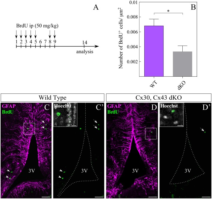
Figure 7.
Loss of gap junction coupling affects hypothalamic cell proliferation. Intraperitoneal injection of bromodeoxyuridine (BrdU) was performed as described in (A). (B) Number of BrdU-positive cells in the parenchyma was quantified in three dKO (13 slices) and four wild type mice (23 slices) and the results were normalized to the area analyzed (μm2), using the ROI manager of ImageJ software. Statistical analysis was performed using the non-parametric Mann Whitney one tail test, *P < 0.05. Representative immunohistochemistry using anti-BrdU (green) and anti-GFAP (magenta) antibodies in frontal sections of WT (C,C′) and dKO (D,D′) animals. Boxes in (C′,D′) indicate co-localization of anti-BrdU with nuclei staining (Hoechst). Arrowheads indicate BrdU staining close to the 3V, while arrows indicate BrdU-positive cells distal to the 3V. 3V, third ventricle. Scale bar (C,C′,D,D′): 50 μm, Scale bar (Hoechst box): 10 μm.

