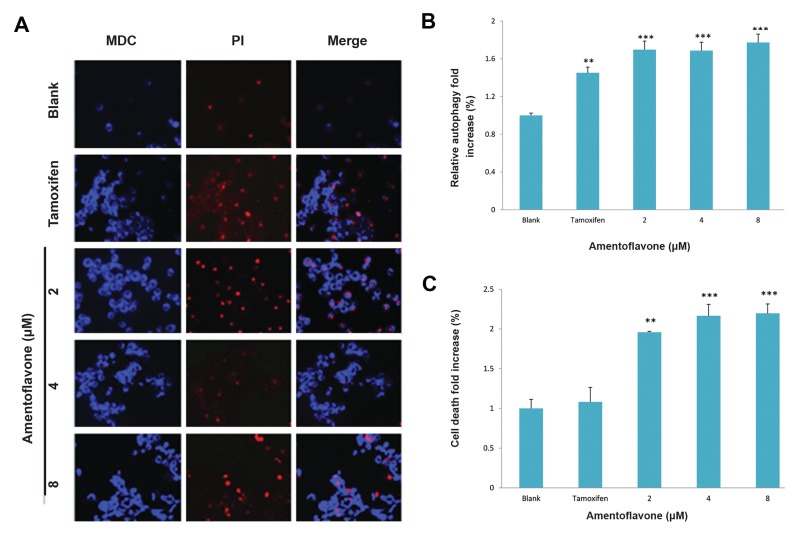
Fig.2.
Effect of amentoflavone on formation of autophagosome in A549 cells. The cells (1×10⁵ cells) were treated with amentoflavone at the indicated concentration. The level of autophagosome formation was evaluated in the presence of amentoflavone or tamoxifen. A. The autophagosome was stained by MDC and the damaged cells or dying cells were stained by PI, B. The effect of amentoflavone on autophagy was analyzed by the fluorescence measurement of autophagic vacuole. The cells showing autophagic vacuoles were quantified by fold increase in green detection reagent signal, and C. The effect of amentoflavone on the cell viability was analyzed by the fluorescence measurement of dead cells stained by propidium iodide. Data are shown as the mean of values ± SD obtained from three independent experiments. The level of significance was identified statistically (**; P<0.01, ***; P<0.001) using Student’s t test. MDC; Monodansylcadaverine and PI; propidium iodide.

