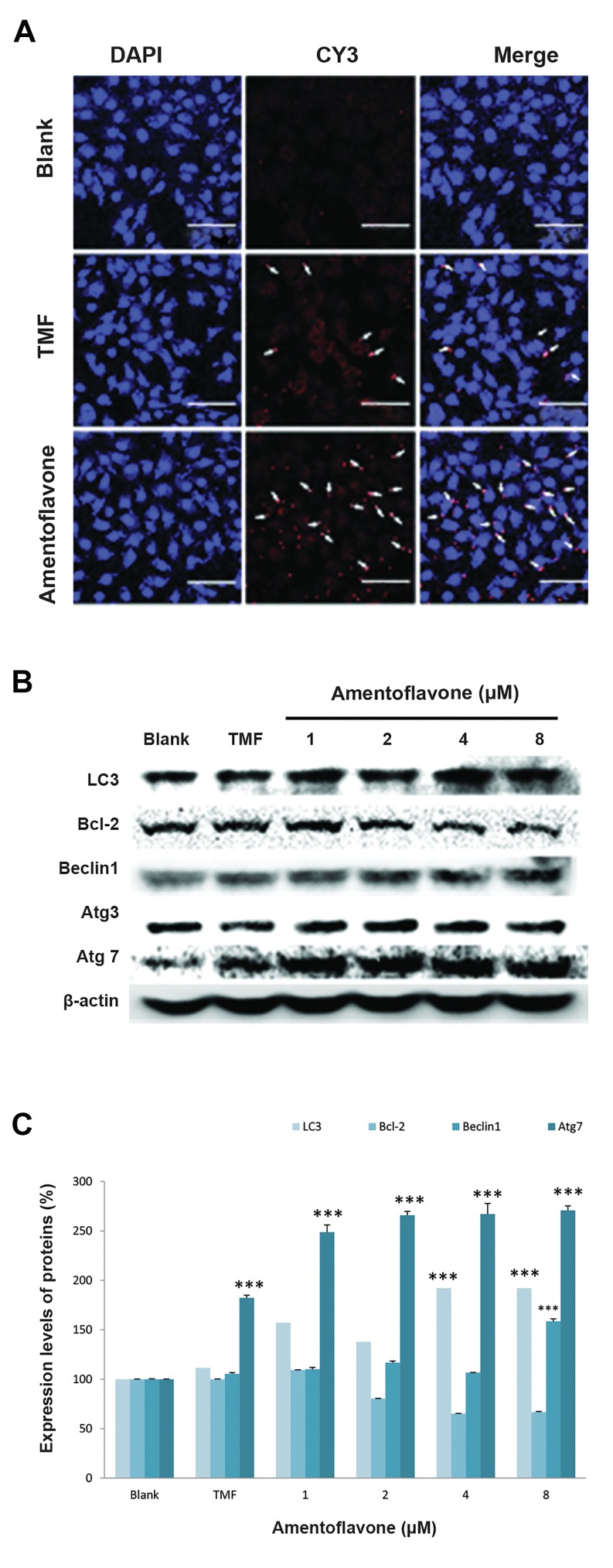
Fig.3.
Effect of amentoflavone on the expression of Atg7 and autophagy- related proteins. A. The images of Atg7 immunofluorescence-stainedA549 cells were shown by the red color. The arrows show Atg7 hasbeen localized to the cell cytosol (scale bar: 100 µm), B. The effect of amentoflavone on protein expressions of LC3, Bcl-2, Beclin1, Atg3, Atg7, and ß-actin was analyzed by western blot, and C. The level of proteins expression was quantified by Multi Gauge V3.0 software. Dataare presented as the mean of values ± SD from three independentexperiments. The level of significance was identified statistically (***; P<0.001) using Student’s t test.

