Fig. 4.
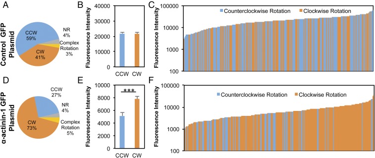
Directional bias of rotational behavior by individual cells can be regulated by α-actinin-1. (A and D) MDCK cells transiently transfected with pEGFP-N1 α-actinin-1 (D) exhibited a predominantly CW rotation, in contrast to those transfected with the control plasmid, pEGFP-N1 (A). (B and E) Quantification of the total EGFP fluorescence intensity per cell reveals that the CW-rotating MDCK cells exhibited greater average expression of α-actinin-1 compared with the cells identified with CCW rotational motion (E), while there was no significant difference between CW- and CCW-rotating cells transfected with the control plasmid (B). (C and F) Histograms displaying the distribution of CCW and CW cells transfected with pEGFP-N1 (C) or pEGFP-N1 α-actinin-1 plasmids (F) with the increase in fluorescence intensity. Each bar represents an individual CW (orange) or CCW (blue) cell, sorted in ascending order of EGFP fluorescence expression. Cells expressing higher levels of α-actinin-1 expression rotated mostly CW, while there were nearly equal instances of CW and CCW rotation in the cells with lower α-actinin-1 expression. MDCK cells transfected with the control plasmid exhibited a slight bias toward CCW, which did not change with changes in fluorescence intensity. ***P < 0.001.

