
Figure 4. PTPσ deletion increases infiltration of IBA-1+ macrophages and CD3+ T cells into spinal cord of EAE mice.
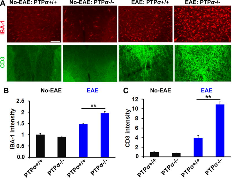
(A) Representative transverse spinal cord sections stained for IBA-1 (red) and CD3 (green) display greater numbers of IBA-1+ macrophages and CD3+ T cells in PTPσ−/− EAE mice. (B, C) Graphs indicate quantification of IBA-1 and CD3+ immunostaining signals from the spinal cord of no-EAE and EAE mice. Scale: 50 μm. n = 3–5 mice/group.