
Figure 9. PTPσ deficiency promotes differentiation of CD4+ T cells into Th1 and Th17 cells.
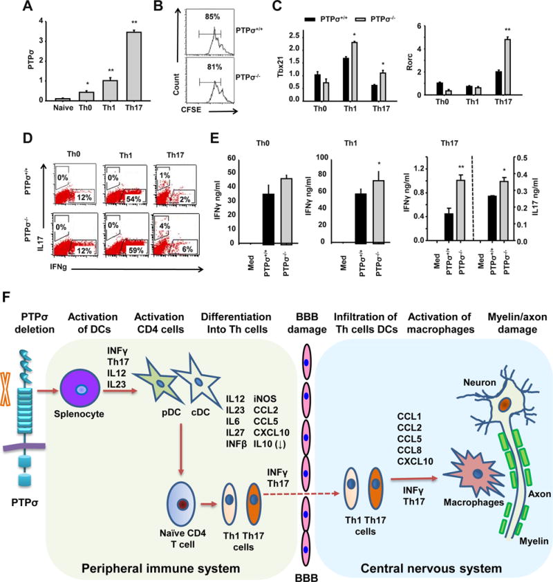
(A, C) Naïve CD4+ T cells purified from spleens of PTPσ +/+ and −/− mice were activated with anti-CD3 (3μg/ml) and anti-CD28 (1μg/ml) in Th0 (no cytokines were added), Th1 (+IL12 20 ng/ml) and Th17 (TGFb 3 ng/ml, IL23 50 ng/ml, IL6 20 ng/ml) conditions for 2 days. The expression levels of PTPσ (A), Tbx21 and Rorc (C) were measured by qRT-PCR. (B) Naïve CD4+ T cells purified from spleens of PTPσ +/+ and −/− mice were activated with anti-CD3 (3μg/ml) and anti-CD28 for 3 days and their proliferation then measured by carboxyfluorescein succinimidyl ester dilution. (D, E) Naïve CD4+ T cells purified from spleens of PTPσ +/+ and −/− mice were activated with anti-CD3 (3μg/ml) and anti-CD28 (1μg/ml) in Th0 (no cytokines), Th1 (+IL12 20 ng/ml) and Th17 (TGFb 3 ng/ml, IL23 50 ng/ml, IL6 20 ng/ml) conditions for 3 days. The levels of cytokines were measured by flow cytometry and ELISA. * p<0.05, ** p<0.01 compared with PTPσ +/+ group. n = 3 independent experiments. (F) Based on our results, we propose a model for the roles of PTPσ in regulating functions of immune cells and development of EAE.