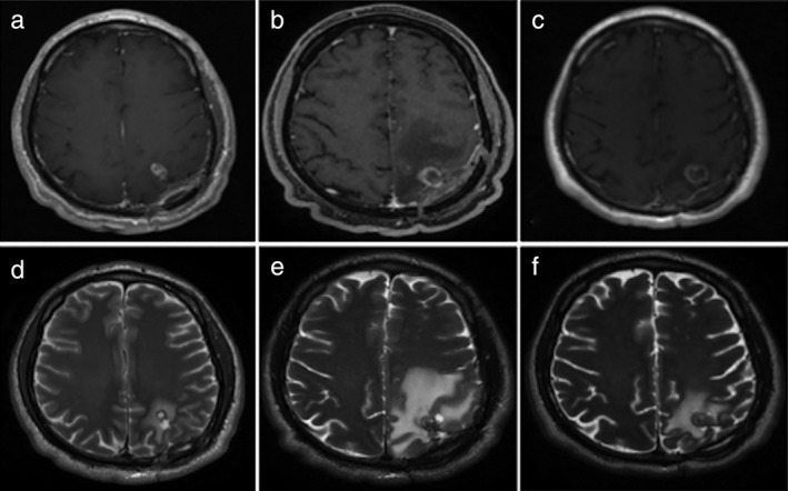
Figure 2.

Magnetic resonance imaging before and after treatment. (a,b) Post‐contrast T1 and T2 weighted images revealed a frontal enhancing solid metastatic lesion with peripheral edema within the brain parenchyma, respectively, prior to initiation of treatment. (c,d) Post‐contrast T1 and T2 weighted images obtained one week after the onset of treatment with nivolumab and ipilimumab revealed an increase in the size and the edema of the metastatic lesion, respectively, with a target‐like appearance. (e,f) Post‐contrast T1 and T2 weighted images obtained eight weeks after the onset of treatment revealed cystic like appearance with thin peripheral enhancement of the metastatic lesion and decrease of the edema, respectively.
