
Figure 7.
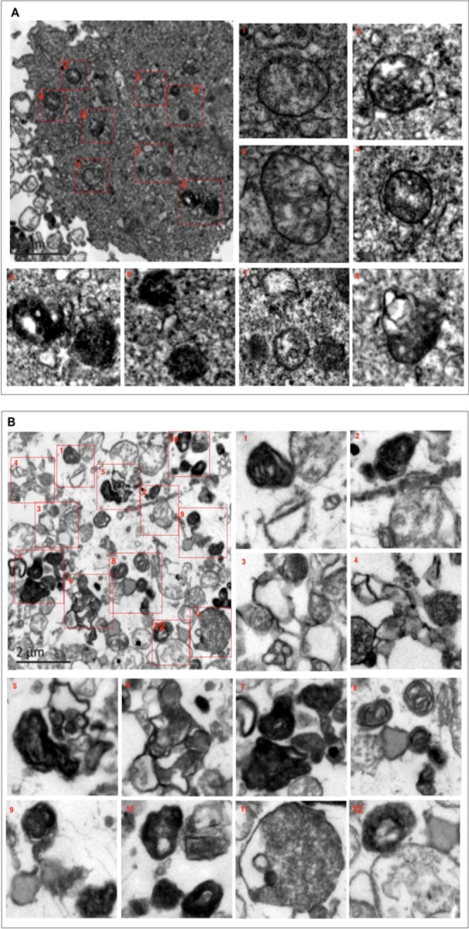
Confirmation of “autophagic stress” induced by combined IADB + 5-FU treatment using TEM: (A) TEM images of HT-29 cells treated with 5-FU (5 μM) for 24 h, revealing minimal autophagic vesicles accompanied by modestly swollen mitochondria with a relatively indistinct double-membrane structure. (B) TEM images of HT-29 cells co-treated with 5-FU (5 μM) + IADB (10 μM) for 24 h, revealing numerous autophagosomes and autolysosomes (electron-dense organelles engulfing double-membraned vesicles). These structures appeared to contain degraded cellular material, and the mitochondria were swollen with crista fragmentation.