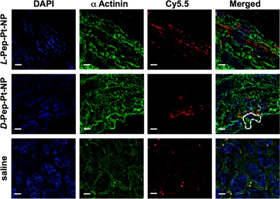
Figure 3.

SIM analysis of 5 μm thick tumor tissue sections of animals pretreated with L-Pep-Pt-NP (top), D-Pep-Pt-NP (middle), or saline (bottom). The samples were examined using specific fluorescent probes. The polymers used to obtain the nanostructures contain a Cy5.5 NIR dye chemically bound to the polymer backbone used to track the nanomaterials (red). Actin filaments were visualized by staining with a mouse anti-α-actinin antibody followed by AF 488 goat antimouse antibody (green). Nucleus was stained with DAPI (blue). Although DAPI is known to bind and stain DNA, some nonspecific binding to other subcellular structures was observed. From these fluorescent images, it is clear that the signal for both the responsive and the nonresponsive nanoparticles are not correlated with nuclear and actin staining, suggesting an extracellular localization of the nanomaterials. Interestingly, samples from animals treated with saline solution showed fluorescent signals in the far red channel, attributed to tissue autofluorescence. The dashed line outlines a cell boundary. Scale bar represents 5 μm.
