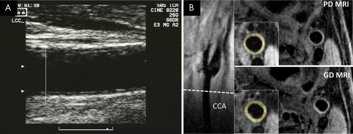
Figure 2:
Images show common carotid artery (CCA) wall thickness assessment in a 62-year-old man by using, A, US and, B, MRI. Far wall intima-media thickness was measured below carotid bulb, starting at point where outer wall of artery begins to diverge (A). Corresponding MR images (B) show location of CCA section (dashed line) on long-axis view of left carotid bifurcation, with noncontrast proton-density (PD)–weighted and intravenous gadolinium-enhanced (GD) MR images obtained at this location. Insets show inner and outer boundaries of segmented vessel walls, with white radial lines delineating 12 sectors over which wall thickness was computed.

