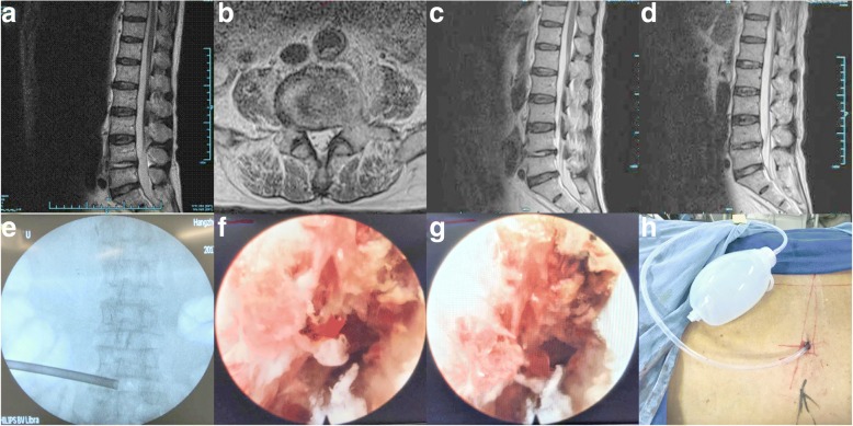
Fig. 1.
Typical case (case number 2). A 71-year-old man was diagnosed with having L5–S1 infectious spondylitis. A sagittal and axial T2-weighted MRI revealed L5–S1 infection with a paraspinal abscess (a, b). Unilateral percutaneous endoscopic debridement and drainage was performed (e, h). On endoscopic views, after the necrotic tissue and pus were discharged, the chapped disc was evident (f, g). Postoperative sagittal T2-weighted MRI at the 1 week (c) and 1 month (d) follow-ups demonstrated a decrease in the abnormally high signal in the L5/S1 intervertebral disc and a disappearance of the abscess.

