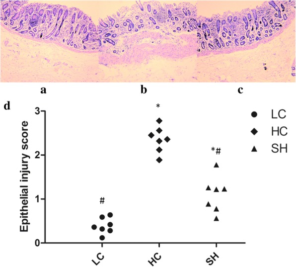
Fig. 2.

Histological alteration of the colonic epithelium of goats from the different groups. Representative photomicrographs with haematoxylin and eosin staining. The colonic epithelium of the LC group was intact and showed no disruption (a), while the stratum corneum structure of the epithelium was severely damaged in the HC group (b). Slight damage of the epithelium was observed in the SH group (c). Epithelial injury scores are shown in d. * indicates P < 0.05 and ** indicates P < 0.01 compared with the LC; # indicates P < 0.05 compared with the HC. A statistically significant difference has a P value < 0.05
