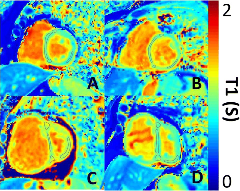
Fig. 2.

Representative T1 maps. Representative T1 maps in short axis geometry of a) a healthy subject, b) a patient without pulmonary artery hypertension, c) a patient with idiopathic PAH and d) a patient with left heart diseaes. Demonstrative regions of interest are places on the RV insertion points, interventricular septum and LV free wall
