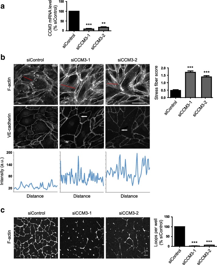
Fig. 2.
CCM3 regulates stress fibers and angiogenic loop formation in endothelial cells. HUVECs were transfected with siRNAs targeting CCM3 or with a control siRNA. a After 72 h, the amount of CCM3 mRNA was determined by qPCR. Data are normalised to GAPDH mRNA levels and are the mean of 3 independent experiments ± SEM. b Left panels, HUVECs were seeded onto fibronectin-coated glass coverslips to form confluent monolayers. 72 h after transfection, cells were fixed and stained for F-actin and VE-cadherin. Images are compressed stacks of 10–15 confocal z-sections. Arrows indicate discontinuous junctions (VE-cadherin); Scale bar, 40 μm. Intensity profiles are indicated for representative cells with the region scanned indicated as a red line on each image. Right panel, stress fibers were quantified as described in Materials and Methods; data show mean ± SEM; n = 3 independent experiments. At least 150 cells were scored per condition in each experiment. c Left panels, 48 h after transfection, HUVECs were seeded onto a layer of Matrigel. Loops were allowed to form for 24 h, and then cells were fixed and stained for F-actin with phalloidin-Alexa546. Scale bar, 100 μm. Right, loop formation was quantified by scoring the number of loops per field using fluorescence images. 6 fields were scored per condition in each experiment. Results are shown as % of siControl. Data are mean ± SEM, n = 3 independent experiments; **p < 0.01, ***p < 0.001 compared to siControl determined by Student’s t-test

