Fig. 6.
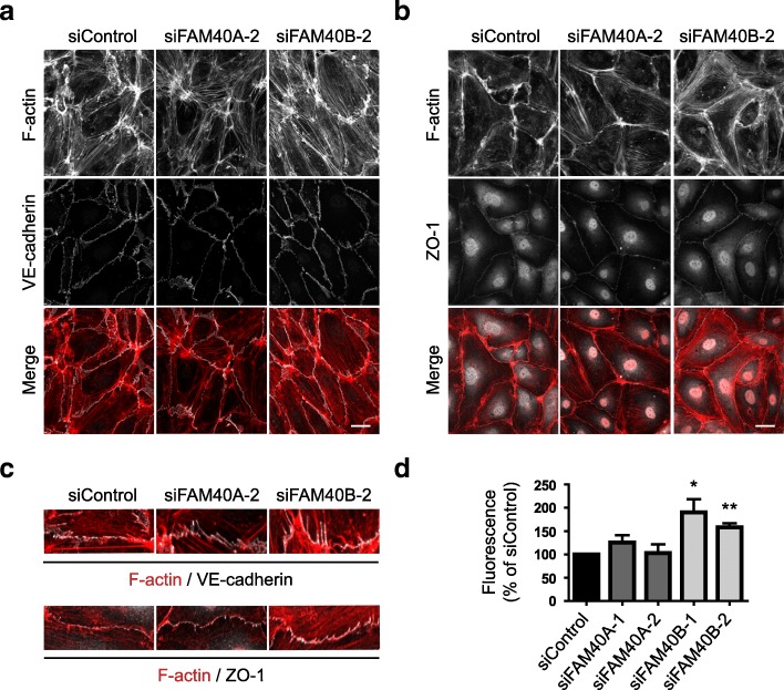
Effect of FAM40A and FAM40B depletion on endothelial junctions and permeability. a HUVECs were transfected with siRNAs targeting FAM40A or FAM40B and seeded onto fibronectin-coated glass coverslips to form monolayers. 72 h after transfection cells were fixed and stained for F-actin and VE-cadherin. b Immunofluorescence analysis of FAM40A and FAM40B-depleted HUVECs for F-actin and ZO-1. Images are compressed stacks of 10–15 z-sections and are representative of 3 independent experiments. Scale bars, 40 μm. c Magnified images highlighting anchoring of stress fibers at cell-cell junctions. d HUVECs were transfected with the indicated siRNAs and plated at confluency onto transwell inserts. 72 h after transfection, FITC-dextran was added to the upper chamber. After a further 80 min the fluorescence of media in the lower chamber was determined. Data show mean permeability ± SEM as % of siControl; n = 3 independent experiments for siFAM40A; n = 5 independent experiments for siFAM40B. *p < 0.05, **p < 0.01; Student’s t-test, compared to siControl

