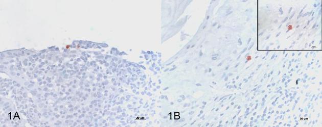
Figure 1.

Immunohistochemistry using a Chlamydiaceae-family-specific mouse monoclonal antibody targeting the chlamydial lipopolysaccharide (LPS, Clone ACI-P, Progen), AEC/peroxidase method, hematoxylin counterstain. (A) Positive immunolabeling of 2 chlamydial inclusions in the epithelium of the rectal mucosa from patient 2 (magnification 400×). (B) Positive immunolabeling of a single chlamydial inclusion in the appendix tissue from patient 4 (magnification 400×). Inset: granular appearance of the chlamydial inclusion in B (magnification 1000×, oil immersion).
