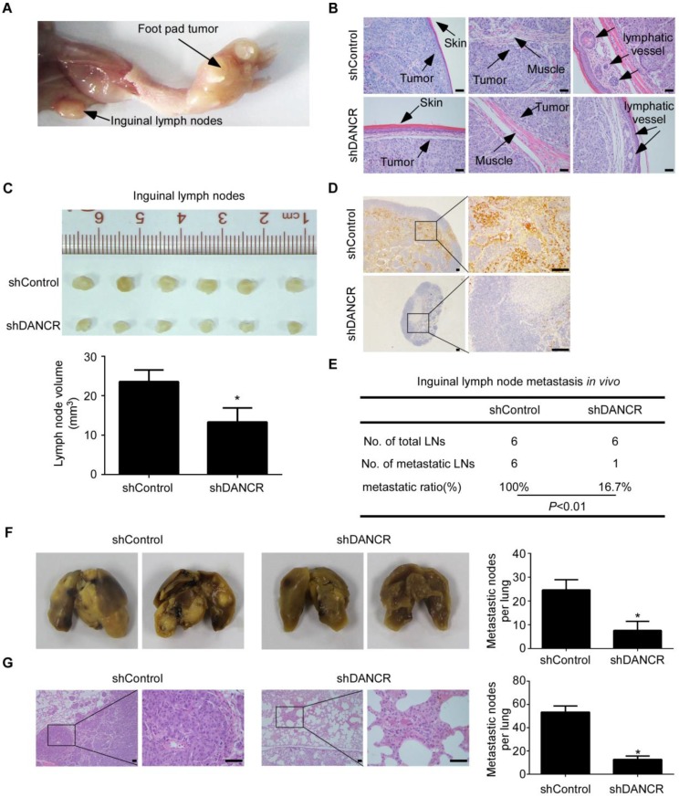
Figure 6.
LncRNA DANCR promotes NPC cell invasion and metastasis in vivo. (A-E) SUNE-1 cells stably expressing shDANCR or control shRNA were transplanted into the foot pads of nude mice (n=6 per group) to construct an inguinal lymph node metastasis model. (A) Representative image of primary tumor in foot pads and metastatic inguinal lymph node. (B) Representative images of microscopic primary foot pad tumors (×200) stained with H&E. Scale bar, 50 μm. (C) Representative image (upper panel) and quantification (lower panel) of the volumes of the inguinal lymph nodes. Data are presented as mean ± SD; Student's t-tests; *P<0.05. (D) Representative images of immunohistochemistry (IHC) staining with pan-cytokeratin in inguinal lymph nodes (left panel ×40, right panel ×200). Scale bar, 100 μm. (E) Inguinal lymph node metastatic ratios. Chi-square test. (F-G) SUNE-1 cells stably expressing shDANCR or control shRNA were injected into the tail veins of nude mice (n=5 per group) to construct a lung metastasis colonization model. (F) Representative images and quantification of macroscopic metastatic nodules on the lung surfaces. Data are presented as mean ± SD; Student's t-tests; *P<0.05. (G) Representative images and quantification of microscopic metastatic nodules in the lung tissues stained with H&E (×100 and ×400). Scale bar, 50 μm. Data are presented as mean ± SD; Student's t-tests; *P<0.05.

