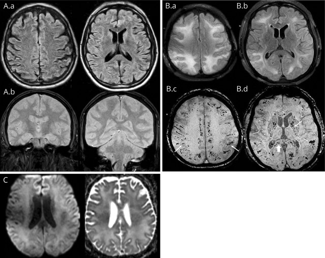
Figure. Imaging.
(A) Axial fluid-attenuated inversion recovery (FLAIR) (A.a) and coronal gradient recalled echo (GRE) (A.b) at presentation to the outside institution show no white matter signal abnormality or hemorrhage. (B) MRI performed 3 weeks after the initial presentation. Axial FLAIR images (B.a, B.b) show extensive confluent hyperintensity in the periventricular, deep, and subcortical white matter. Axial susceptibility-weighted imaging (B.c, B.d) shows numerous microhemorrhages in the subcortical white matter (arrows, B.c), internal capsules (arrow, B.d) and corpus callosum (arrowhead, B.d). (C) MRI performed 3 weeks after the initial presentation. Axial diffusion-weighted imaging and B.a, B.d, and B.c show no restricted diffusion in the white matter. Not pictured is the second MRI FLAIR and GRE obtained at the outside institution prior to transfer, which revealed a similar distribution of leukoencephalopathy and microhemorrhages as the repeat images obtained at our institution and presented in (B).

