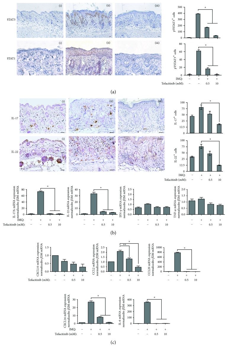
Figure 6.
Tofacitinib counteracts IMQ effects in mouse skin. Immunohistochemistry analysis of mouse skin left untreated (i), IMQ-treated (ii), and IMQ-treated in the presence of tofacitinib (iii) shows reduction of STAT1-, STAT3-, IL-17A-, and IL-22-positive cells (a) and (b) after tofacitinib treatment. Sections were counterstained with Mayer's hematoxylin and were visually evaluated by a pathologist experienced in dermatology. Bars, 200 μM. One of four representative stainings is shown. Graphs show the mean number of positive cells ± SD per three sections per experimental group (n = 10 mice). ∗p < 0.01. In (b), graphs show real-time PCR analyses of IL-17A, IL-22, IFN-γ, and TNF-α performed on pooled mRNA samples (n = 10) of mouse skin treated as indicated. ∗p < 0.01. In (c), graphs show real-time PCR analyses of CXCL10, CCL2, CCL20, CXCL16, and IL-6 performed on pooled mRNA samples (n = 10) of mouse skin treated as indicated. ∗p < 0.01, ∗p < 0.05.

