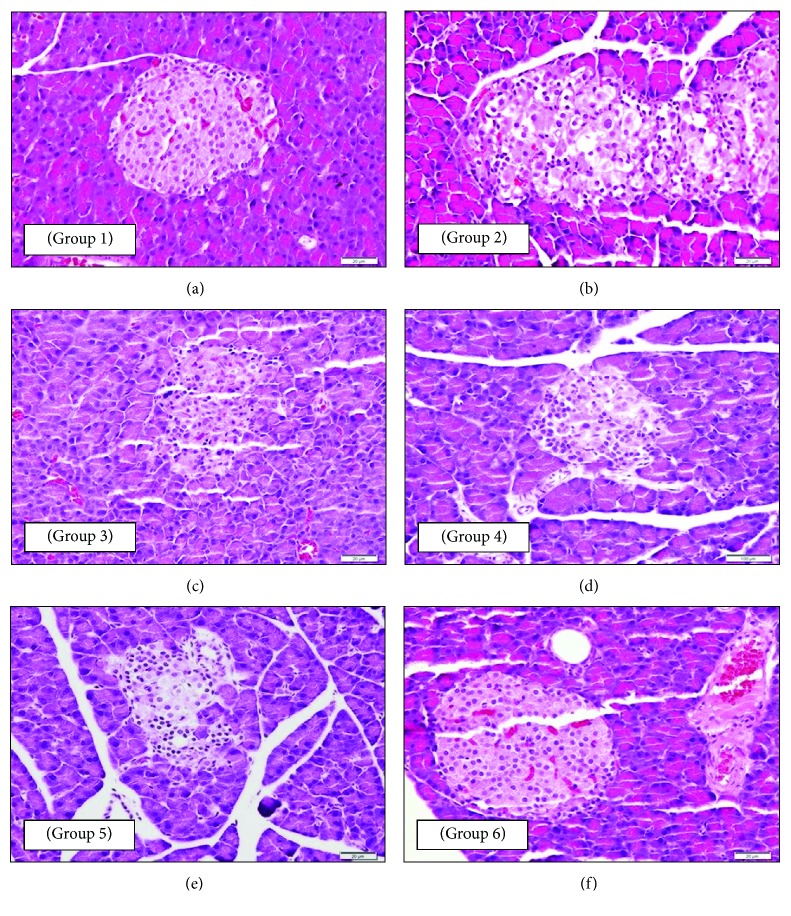
Figure 1.
Photomicrographs of a section of the endocrine pancreatic tissue of rats: (a—group 1: NC) normal pancreatic structure and exocrine acini surrounding the islets of Langerhans (400x magnification); (b—group 2: DC) disorganized islets of Langerhans and clusters of inflammatory cells (β-cells show an unstained vacuolated cytoplasm and dark-stained degenerated nuclei) (400x magnification); (c—group 3: DM) normal islets of Langerhans and blood sinusoids (100x magnification); (d—group 4: DO) islets of Langerhans with a normal-looking population of β-cells (100x magnification); (e—group 5: DOM) normal islets of Langerhans with a normal population of β-cells and absence of any degenerative change (400x magnification); (f—group 6: DOP) normal islets of Langerhans with a normal β-cell population similar to that of group 1 (NC) indicating protection from the damaging effect of STZ (400x magnification). All sections were stained with H and E stain and viewed with a light microscope.

