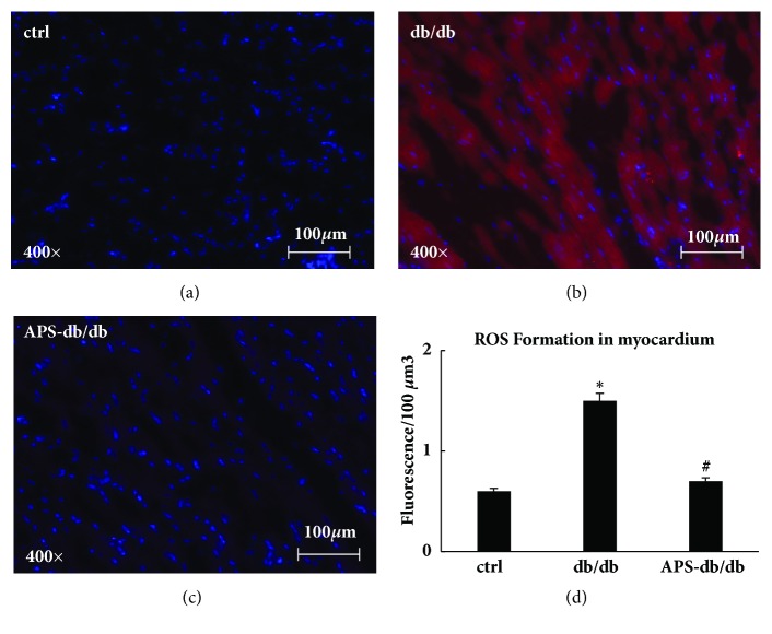
Figure 5.
APS inhibited ROS formation in myocardium of db/db mice. Tissue sections of the left ventricle myocardium were obtained from C57BJ/6J mice and db/db mice with or without APS treatment (n=6 per group). CM-H2DCFDA was employed to measure the ROS formation, and the myocardial H2O2 and ·OH concentration were analyzed utilizing InSpeak Microscopy Image Intensity Calibration Microspheres and ImagePro analysis software. (a)~(c) The fluorescence microphotographs showing ROS formation in myocardium (red: fluorescence for H2O2 and ·OH; blue: cardiomyocytes). (d) ROS formation in myocardium from left ventricle. Values were presented as mean ± standard error of the mean (SEM). ∗P<0.05 versus C57BJ/6J control mice, and #P<0.05 versus db/db mice.

