Figure 5.
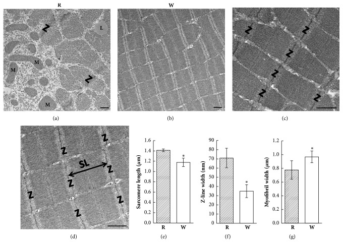
Electron microscopic (EM) ultrastructural images of red (R) and white (W) muscle fibers. (a) Several mitochondria are located near the sarcolemma. Lipid droplets can be seen between the myofibrils in red muscle fibers. (b) EM image indicating less cytoplasm and fewer mitochondria between myofibrils in white muscle fibers. (c) A longitudinal section displayed the well-organized sarcomere in red muscle fibers. (d) Well-organized sarcomeres were also observed in white muscle fibers. The sarcomere length (double-headed arrow) is the distance between two Z-lines. (e) The sarcomere length was shorter in white muscle fibers. (f) The Z-line width was thinner in white muscle fibers. (g) The myofibril width was larger in white muscle fibers. ∗ p < 0.001. L, lipid droplet; M, mitochondria; SL, sarcomere length; Z, Z-line. Bar: 0.5 μm.

