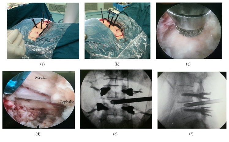
Figure 1.
(a) Percutaneous transforaminal puncture into disk after percutaneous pedicle screw fixation. (b) Sequential dilation. (c) Optional foraminoplasty and expansion of the safety triangle by bone drill under endoscopic views. (d) Neurological decompression and initial endplate preparation in endoscopic view. (e) and (f) Working tube insertion in anteroposterior and lateral X-ray views.

