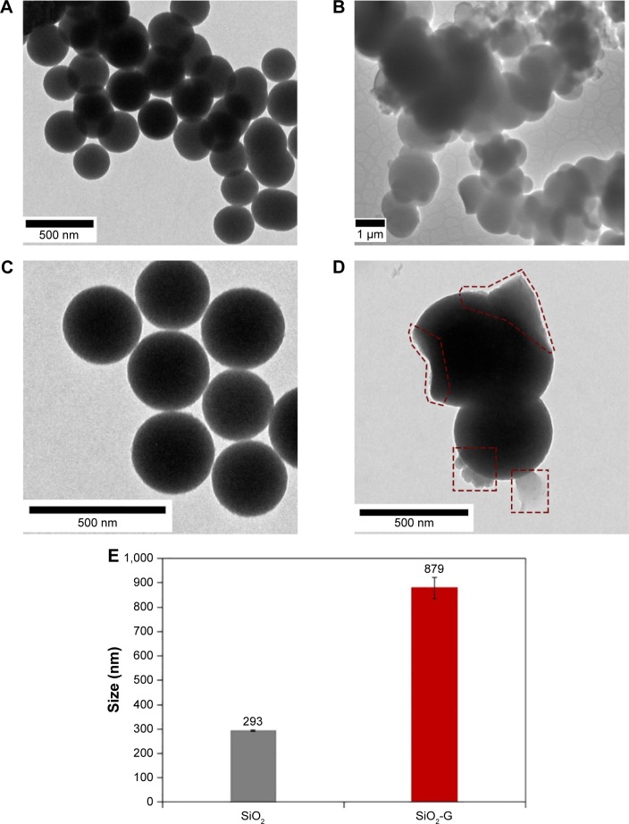
Figure 2.
TEM images of the materials.
Notes: (A, C) Pristine SiO2 NPs and (B, D) SiO2-G nanohybrids. The surface-loaded gentamicin is marked red with dashed shapes in panel D. Magnifications of 8,000× (A), 60,000× (B), and 10,000× (C, D). (E) The size of the pristine SiO2 and SiO2-G nanohybrids analyzed on the obtained TEM, and SEM and TEM images, respectively. The error bars represent the standard errors of the means.
Abbreviations: SEM, scanning electron microscope; SiO2 NPs, silica nanoparticles; SiO2-G, silica–gentamicin; TEM, transmission electron microscope.

