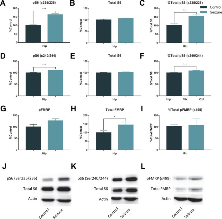
Figure 5.

Results of 1 h western blots for pS6, S6, pFMRP, and FMRP. In samples taken 1 h (control = 9; seizure = 9) after a single seizure there was a significant increase in hippocampal pS6 (s235/236) (A), no difference in levels of hippocampal total S6 (B), and a significant increase in the ratio of pS6 (s235/236)/total S6 (C). There was also a significant increase in pS6 (s240/244) (D), but no difference between groups in total S6 (E), and a significant increase in the ratio of pS6 (s240/244) to total S6 (F). G, There was a trending, but nonsignificant increase in pFRMP. However, there was a significant increase in total FMRP 1 h after a seizure (H), and the ratio of pFMRP to total FMRP was no different between groups (I). J‐L, Representative western blots. The bars represent the mean and the error bars indicate SEM. * p < 0.05, *** p < 0.001.
