
Figure 3.
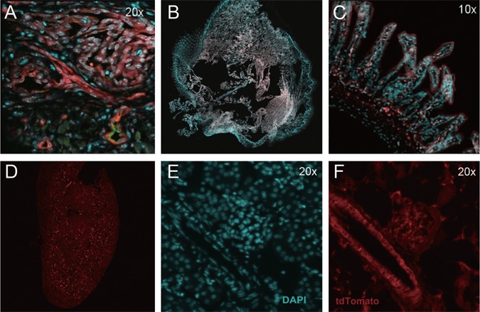
Recombination in pericytes in Tsc2 ff FoxD1 GC+ tdTomato + mice. Recombination, as assessed by tdTomato fluorescence, can be seen in a tongue HPC (A), intestinal wall pericytes (C) and pericytes from kidney blood vessels and glomeruli (D–F). Tongue from control mouse (B, Tsc2fw FoxD1GC+) showed no tdTomato fluorescence, indicating absence of recombination. Red reflects dTomato fluorescence (A, B, C, D, F), cyan indicates DAPI labeled nuclei (A, B, C, E), and the green counterstain in the tongue is endomucin (A, EC marker). Note that A-C are fused images, while D shows the whole image of kidney by tdTomato alone, E shows DAPI signal alone, and F tdTomato fluorescence alone.