
Figure 3.
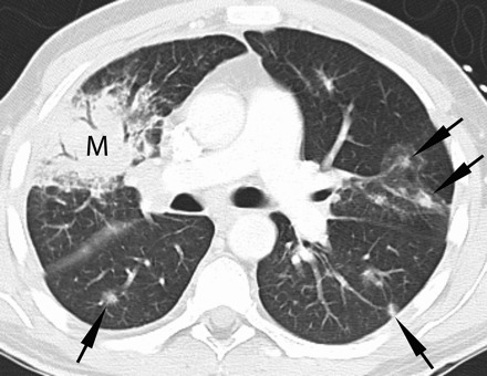
Thirty-eight-year-old man with acute myeloid leukemia, cord blood transplanted with refractory neutropenic fever. Contrast-enhanced chest computed tomography (lung windows) at the level of the main pulmonary artery demonstrates multiple pulmonary nodules (arrows), some of which have a ground glass halo surrounding them and a right upper lobe consolidative mass (M). Biopsy demonstrated fungal hyphae consistent with mucormycosis. Numerous additional nodules were seen throughout the remainder of the lungs (not shown).