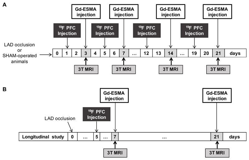
Figure 1.
Experimental study design. Myocardial infarction was induced in C57Bl6 female mice after permanent occlusion of the left anterior descending coronary artery (LAD). MRI scans were performed after intravenous injection of 19F PFCs and Gd-ESMA, 48 and 1h before imaging sessions, respectively. (A) Mice (n=8 per group/time point) were imaged at 3, 7, 14 and 21 days post-MI. SHAM-operated mice (n=6 per group/time point) were imaged at the same time-points and were used as controls. At the end of the scans, mice were culled and hearts were extracted for histology and NMR (n=4/group and 3-SHAM-operated animals/time-point for each technique) (B) 15 mice were imaged longitudinally at 7 and 21 days post-MI. MRI, magnetic resonance imaging; 19F PFC, 19F perfluoro-15-crown-ether emulsions; Gd-ESMA, elastin/tropoelastin specific MR contrast agent.

