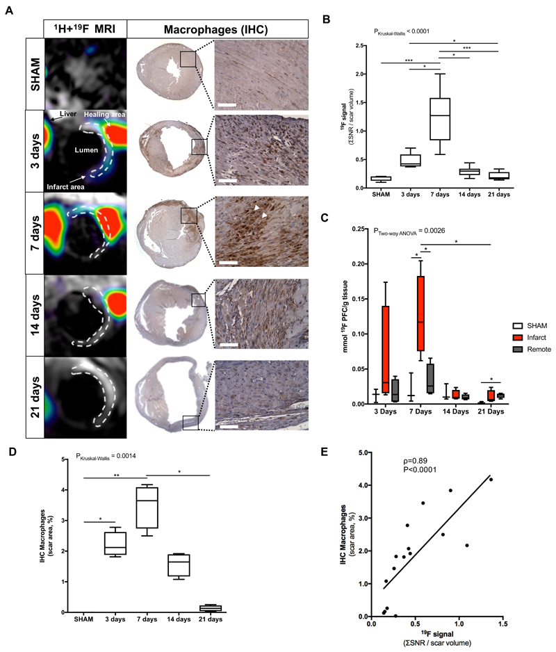
Figure 3.
Assessment of inflammatory response after myocardial infarction in mice at 3T MRI using 19F perfluorocarbons. (A) Representative short-axis views of co-registered 1H+19F images (left column; N=8 MI animals/time-point, N=6 SHAM-operated animals/time-point) and macrophage IHC (macrophages identified as MAC-3 positive, brown. N=4 MI animals/timepoint, N=3 SHAM-operated animals/time-point) from the heart at 3, 7, 14 and 21 days after MI. (B) In vivo 19F MRI signal quantification. (C) Ex vivo 19F NMR signal quantification (N=4/timepoint, N=3 SHAM-operated animals). (D) IHC macrophages quantification shown a significantly decrease between 7 and 21 days after infarct. (E) Correlation between ex vivo macrophages IHC and in vivo 19F MRI signal. Spearman correlation (N=20, ρ=0.89, P<0.0001). IHC:immunohistochemistry. Scale bar, 50μm. *P<0.05,**P<0.01,***P<0.001.

