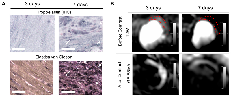
Figure 4.
Assessment of elastin/tropoelastin and myocardial edema in mice with myocardial infarction. (A) Tropoelastin IHC and Elastica van Gieson (black staining) reveal the absence of protein deposition within the heart at 3 days, starting to accumulate at day 7 (arrows). (B) T2-weighted images showing high signal intensity on the lateral wall (top images); Contrast-enhanced image (late gadolinium enhancement) showing high signal intensity in the infarcted areas (middle images). IHC:immunohistochemistry. Scale bar, 25μm.

