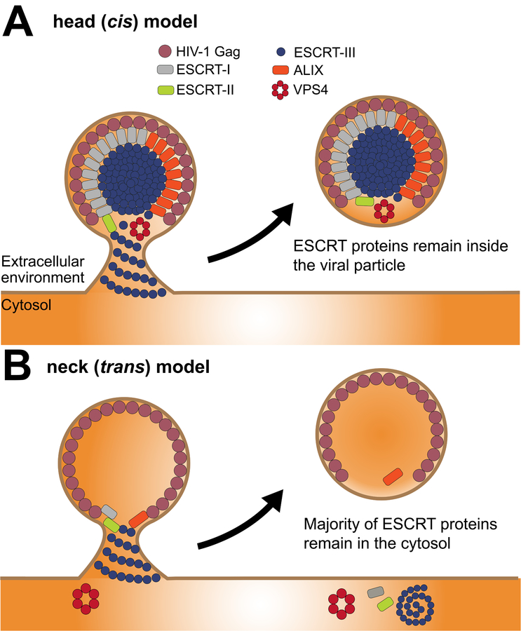
Figure 2. ESCRT topology in HIV-1 viral particle release.
An overview of the proposed HIV-1 Gag and ESCRT interactions during the viral release mechanism. (A) The cis model (cis with respect to the contents of the viral bud) postulates that ESCRTs form assemblies and sever the neck from within. This could be driven very locally from the vicinity of the neck (not shown), or in the model shown here, from the head of the virion. The cis mechanism must result in at least some ESCRT proteins departing with the viral particle when it liberated from the PM, but the number of molecules need could be small enough to be difficult to detect. The head-driven version shown here would lead to large amounts of ESCRTs incorporated into viral particles, which does not fit most of the data in the field, but does have its advocates (65). (B) In comparison with the cis model, the trans model or neck model shows that ESCRT subunits polymerize and recruit VPS4 at the neck where membrane abscission is achieved. Finally, ESCRTs are recycled back to the cytosolic pool although substantial amounts of ALIX and trace amounts of other ESCRTs may be found present inside the virion.

