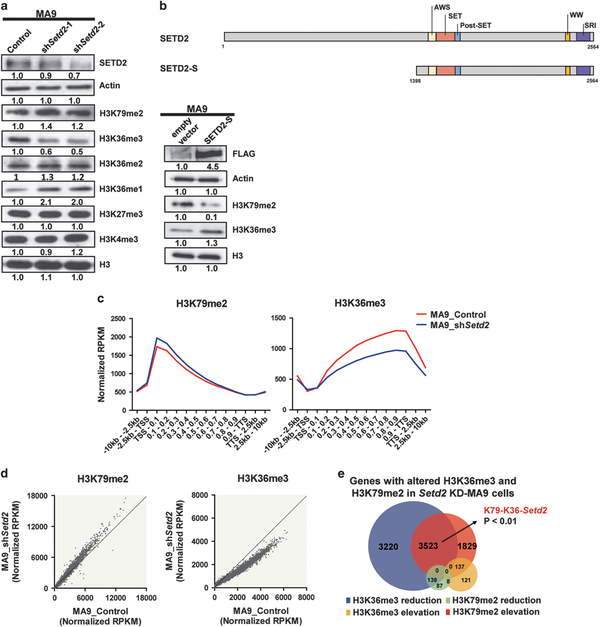
Figure 3.
SETD2-H3K36me3 loss-of-function further elevated H3K79me2. (a) Immuno-blotting results after Setd2 KD in MA9 cells. (b) Schematic diagram showing major functional domains in SETD2 and the constructed SETD2-S vector. Immuno-blotting of SETD2-S and empty vector expression in MA9 cells. (c) ChIP-seq profiles of H3K79me2 and H3K36me3 for gene bodies (left: **P<0.01; right: ***P<0.001) in control (MA9_Control) and Setd2 KD-MA9 cells (MA9_shSetd2). (d) H3K79me2 and H3K36me3 ChIP signals on each gene. (e) Venn diagram of genes with altered H3K79me2 and/or H3K36me3 occupancies at gene bodies after Setd2 KD in MA9 cells. See also Supplementary Table S1.

