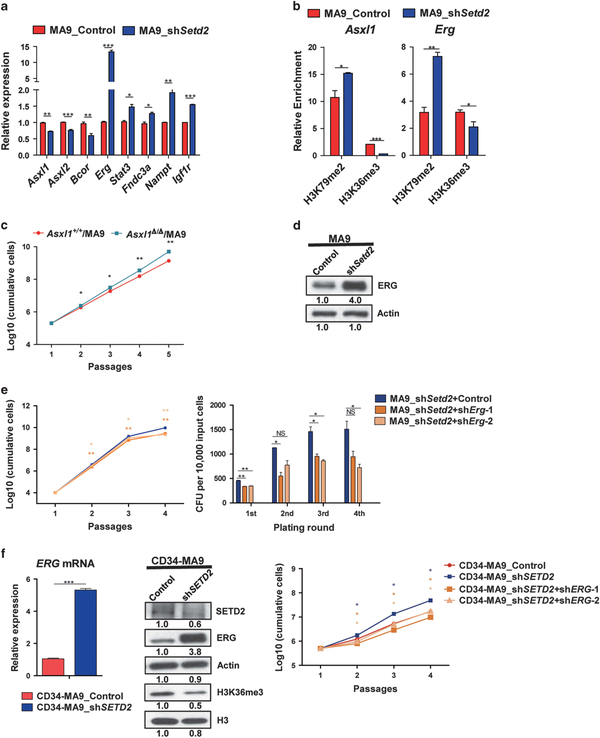
Figure 5.
Both downregulation of Asxl1 and upregulation of Erg/ERG contribute to Setd2/SETD2 KD-mediated leukemia acceleration. (a) DEGs in Setd2 KD-MA9 cells were validated by RT-PCR. P-values were calculated by two-tailed Student’s t-test (*P<0.05, **P<0.01, ***P<0.001). Data are represented as mean ± s.d. (n = 2). Representative data are shown. (b) ChIP-qPCR of H3K79me2 and H3K36me3 at Asxl1 and Erg gene bodies. Enrichment was normalized to input samples (*P<0.05). Data are represented as mean ± s.d. (n = 2). (c) Enhanced proliferation of Asxl1Δ/Δ/MA9 cells compared to Asxl1+/+/MA9 cells was observed. P-values were calculated by two-tailed Student’s t-test (*P<0.05, **P<0.01). Data are represented as mean ± s.d. (n = 2). (d) Overexpression of ERG after Setd2 KD in MA9 cells was detected by immuno-blotting. (e) Decreased proliferation and self-renewal capacities in Erg KD MA9_shSetd2 cells (MA9_shSetd2+shErg) compared to control cells (MA9_shSetd2 +Control) (*P<0.05, **P<0.01). Data are represented as mean ± s.d. (n = 2). (f) Activation of ERG after SETD2 KD in human CD34-MA9 cells (CD34-MA9_shSETD2) compared to control cells (CD34-MA9_Control) (***P<0.001) was detected by RT-PCR and immuno-blotting. Enhanced cell proliferation of CD34-MA9_shSETD2 cells compared to CD34-MA9_Control cells was observed. ERG KD decreased cell proliferation in CD34-MA9_shSETD2 cells (CD34-MA9_shSETD2+shERG) compared to control cells (CD34-MA9_shSETD2) (*P<0.05).

